Product info

BHQ™ Probes are traditional, linear, dual-labelled FRET probes, typically 20 to 30 bases in length, with a fluorophore and quencher covalently attached to the 5' and 3' ends, respectively. Fluorescence is released through the 5' exonuclease activity of Taq polymerase, which cleaves off the fluorescent dye upon the probe's hybridisation to its complementary sequence. BHQ Probes are ideal for detecting the presence and quantifying the amount of specific target sequences.

Key benefits:

  • Unmatched quality and technical support from the original makers of the BHQ quenchers
  • Complete suite of fluorescent dyes spanning the entire visible spectrum
  • Simple probe design and implementation with consistent and reliable performance
 

Design versatility without performance variability

When it comes to assay design, we don’t believe in a one-dye-fits-all strategy. Our wide selection of dyes empowers you to design the best PCR or qPCR probe for your application without sacrificing performance. We have paired each dye with the BHQ quencher that offers optimal quenching efficiency. With our versatile BHQ Probes, you can design with confidence.

5' fluorescent dye

Abs (nm)

Em (nm)

3' quencher

  FAM 495 520 BHQ-1
  TET 521 536 BHQ-1
  CAL Fluor Gold 540 522 544 BHQ-1
  CIV-550 530 550 BHQ-1*, BHQ-2
  JOE 529 555 BHQ-1
  HEX 535 556 BHQ-1
  CAL Fluor Orange 560 538 559 BHQ-1
  Quasar 570 548 566 BHQ-2
  Cy3 549 566 BHQ-2
  TAMRA 557 583 BHQ-2
  CAL Fluor Red 590 569 591 BHQ-2
  ROX 586 610 BHQ-2
  CAL Fluor Red 610 590 610 BHQ-2
  CAL Fluor Red 635 618 637 BHQ-2
  Cy5 646 669 BHQ-2
  Quasar 670 647 670 BHQ-2*, BHQ-3
  Quasar 705 690 705 BHQ-2*, BHQ-3

* Recommended quencher

Use the spectral overlay tool to view compatible dyes for your instrument. Learn more

High quality FAM/BHQ-1 ValuProbes at a low price

Our ValuProbe is comprised of 10 nmols probe with a 5' FAM and a 3' BHQ-1 quencher, delivered dried down, purified via Reverse Phase HPLC, and verified via mass spectrometry and UHPLC.

How to order a ValuProbe

  • Navigate to the Order now tab at the top of this page.
  • Select   valuprobe   to populate ValuProbe parameters.
  • Enter your probe name and sequence.
  • Add primers (optional).
  • Add to basket.

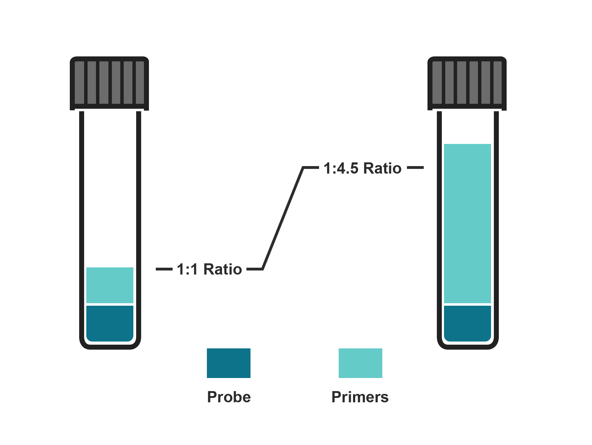
A ValuMix assay built exactly to your specifications

BHQ Probes in the ValuMix assay format for qPCR and gene expression include a BHQ Probe and two primers mixed at your designated primer to probe ratio between 1 and 4.5. Simplify and streamline your experimental setup while reducing any chance of pipetting error.

Key features of ValuMix assays

  • Probes purified via RP HPLC
  • Salt-free primers
  • Wide selection of dyes: FAM, TET, CAL Fluor Gold 540, CIV-550, HEX, CAL Fluor Orange 560, CAL Fluor Red 610 and Quasar 670
  • 0.5 nmol (FAM only), 5 nmol, or 20 nmol delivered

Design considerations for BHQ Probe

  • Target a probe length typically between 20 and 30 bases.
  • If your sequence is shorter than 17 bases, consider ordering this sequence as a BHQplus Probe.
  • BHQ Probe sequences longer than 30 bases are unlikely to have efficient quenching. Consider ordering a BHQnova for sequences 30 bases or longer. For assistance, please contact us.
  • Target a %GC content between 30% and 80%.
  • Avoid runs of identical nucleotides.
  • Do not place a guanosine on the 5′ end. A guanosine next to the reporter dye will alter the fluorescence.
  • Avoid guanosine on the 3′ end (ex. 5′-...GGG-3′ or 5′-...GGAG-3′).
  • Avoid 4 consecutive guanosines as these can form a stable secondary structure.
  • For FAM-labelled probes, avoid a guanosine in the second position on the 5′ end.
  • Building probes with an internal quencher (currently available with FAM only) introduces an additional T base into the sequence.
  • Use IUPAC code when entering wobble/degenerate bases in the Sequence Entry field instead of using parentheses, e.g. (A/G), (A/G/C/T), etc.

Order now

BHQ Probes usually ship in 5 business days.
  • Oligos for North America, South America, and APAC will be supplied out of our Petaluma, US manufacturing facility while oligos for EMEA will be supplied out of our Lystrup, Denmark manufacturing facility unless you have an agreement in place.
  • Our advertised turnaround time applies to orders received before 12PM for the local manufacturing site. Turnaround time is defined as the time of checkout until shipment.

Similar product

BHQ ValuMix assays for qPCR and gene expression offer the convenience of having a custom, dual-labeled BHQ probe and primer pair delivered in exact amounts within a single tube at your selected probe-to-primer ratio (between 1:1 to 1:4.5).


Order BHQ probes and primers delivered each in separate tubes

Oligo set 1

ValuProbe

add
Probe nmol options

Purification
Item price $0.00 TBD

Item price $0.00

Item price $0.00
nmol options
Clear scale

You are ordering:
Probe catalog #: Primer catalog #: Assay subtotal: $0.00 Qty another assay
Note

Subtotal: $0.00 add to cart

Product listing

Catalog # Item name Price Note
DLO-C3B2-1
Dual-labeled Probe, 5' Cy3/3' BHQ-2 $661.00 Provides 50 nmol delivered.
DLO-C3B2-2
Dual-labeled Probe, 5' Cy3/3' BHQ-2 $528.00 Provides 25 nmol delivered.
DLO-C3B2-5
Dual-labeled Probe, 5' Cy3/3' BHQ-2 $397.00 Provides 10 nmol delivered.
DLO-FB1-1
Dual-labeled Probe, 5' FAM/3' BHQ-1 $550.00 Provides 80 nmol delivered.
DLO-FB1-2
Dual-labeled Probe, 5' FAM/3' BHQ-1 $374.00 Provides 35 nmol delivered.
DLO-FB1-5
Dual-labeled Probe, 5' FAM/3' BHQ-1 $223.00 Provides 15 nmol delivered.
DLO-FT-25
Dual-labeled Probe, 5' FAM/3' TAMRA $179.00 Provides 5 nmol delivered.
DLO-FT-1
Dual-labeled Probe, 5' FAM/3' TAMRA $550.00 Provides 80 nmol delivered.
DLO-FT-2
Dual-labeled Probe, 5' FAM/3' TAMRA $374.00 Provides 35 nmol delivered.
DLO-FT-5
Dual-labeled Probe, 5' FAM/3' TAMRA $223.00 Provides 15 nmol delivered.
DLO-HB1-1
Dual-labeled Probe, 5' HEX/3' BHQ-1 $676.00 Provides 80 nmol delivered.
DLO-HB1-2
Dual-labeled Probe, 5' HEX/3' BHQ-1 $463.00 Provides 35 nmol delivered.
DLO-HB1-5
Dual-labeled Probe, 5' HEX/3' BHQ-1 $304.00 Provides 15 nmol delivered.
DLO-HB1-25
Dual-labeled Probe, 5' HEX/3' BHQ-1 $179.00 Provides 5 nmol delivered.
DLO-JB1-1
Dual-labeled Probe, 5' JOE/3' BHQ-1 $903.00 Provides 45 nmol delivered.
DLO-JB1-2
Dual-labeled Probe, 5' JOE/3' BHQ-1 $671.00 Provides 15 nmol delivered.
DLO-RB2-1
Dual-labeled Probe, 5' ROX/3' BHQ-2 $903.00 Provides 45 nmol delivered.
DLO-RB2-2
Dual-labeled Probe, 5' ROX/3' BHQ-2 $671.00 Provides 15 nmol delivered.
DLO-TEB1-1
Dual-labeled Probe, 5' TET/3' BHQ-1 $632.00 Provides 80 nmol delivered.
DLO-TEB1-2
Dual-labeled Probe, 5' TET/3' BHQ-1 $434.00 Provides 35 nmol delivered.
DLO-TEB1-5
Dual-labeled Probe, 5' TET/3' BHQ-1 $297.00 Provides 15 nmol delivered.
DLO-TEB1-25
Dual-labeled Probe, 5' TET/3' BHQ-1 $179.00 Provides 5 nmol delivered.
DLO-TB2-1
Dual-labeled Probe, 5' TAMRA/3' BHQ-2 $603.00 Provides 80 nmol delivered.
DLO-TB2-2
Dual-labeled Probe, 5' TAMRA/3' BHQ-2 $418.00 Provides 35 nmol delivered.
DLO-TB2-5
Dual-labeled Probe, 5' TAMRA/3' BHQ-2 $297.00 Provides 15 nmol delivered.
DLO-TB2-25
Dual-labeled Probe, 5' TAMRA/3' BHQ-2 $179.00 Provides 5 nmol delivered.
DLO-TET-1
Dual-labeled Probe, 5' TET/3' TAMRA $632.00 Provides 80 nmol delivered.
DLO-TET-2
Dual-labeled Probe, 5' TET/3' TAMRA $441.00 Provides 35 nmol delivered.
DLO-TET-5
Dual-labeled Probe, 5' TET/3' TAMRA $297.00 Provides 15 nmol delivered.
DLO-TET-25
Dual-labeled Probe, 5' TET/3' TAMRA $179.00 Provides 5 nmol delivered.
DLO-CAB2-2
Dual-labeled Probe, 5' CAL Fluor Red 610/3' BHQ-2 $461.00 Provides 35 nmol delivered.
DLO-CAB2-25
Dual-labeled Probe, 5' CAL Fluor Red 610/3' BHQ-2 $179.00 Provides 5 nmol delivered.
DLO-CAB2-5
Dual-labeled Probe, 5' CAL Fluor Red 610/3' BHQ-2 $320.00 Provides 15 nmol delivered.
DLO-CAB2-1
Dual-labeled Probe, 5' CAL Fluor Red 610/3' BHQ-2 $715.00 Provides 80 nmol delivered.
DLO-C5B2-5
Dual-labeled Probe, 5' Cy5/3' BHQ-2 $397.00 Provides 3 nmol delivered.
DLO-C5B2-2
Dual-labeled Probe, 5' Cy5/3' BHQ-2 $528.00 Provides 10 nmol delivered.
DLO-C5B2-1
Dual-labeled Probe, 5' Cy5/3' BHQ-2 $766.00 Provides 40 nmol delivered.
DLO-Q6B2-5
Dual-labeled Probe, 5' Quasar 670/3’ BHQ-2 $334.00 Provides 10 nmol delivered.
DLO-Q6B2-2
Dual-labeled Probe, 5' Quasar 670/3’ BHQ-2 $470.00 Provides 25 nmol delivered.
DLO-Q6B2-1
Dual-labeled Probe, 5' Quasar 670/3’ BHQ-2 $715.00 Provides 50 nmol delivered.
DLO-Q6B2-25
Dual-labeled Probe, 5' Quasar 670/3’ BHQ-2 $179.00 Provides 5 nmol delivered.
DLO-Q5B2-5
Dual-labeled Probe, 5' Quasar 570/3’ BHQ-2 $320.00 Provides 15 nmol delivered.
DLO-Q5B2-2
Dual-labeled Probe, 5' Quasar 570/3’ BHQ-2 $470.00 Provides 35 nmol delivered.
DLO-Q5B2-1
Dual-labeled Probe, 5' Quasar 570/3’ BHQ-2 $676.00 Provides 80 nmol delivered.
DLO-Q5B2-25
Dual-labeled Probe, 5' Quasar 570/3’ BHQ-2 $179.00 Provides 5 nmol delivered.
DLO-Q6B3-2
Dual-labeled Probe, 5' Quasar 670/3’ BHQ-3 $470.00 Provides 25 nmol delivered.
DLO-Q6B3-1
Dual-labeled Probe, 5' Quasar 670/3’ BHQ-3 $715.00 Provides 50 nmol delivered.
DLO-Q6B3-5
Dual-labeled Probe, 5' Quasar 670/3’ BHQ-3 $341.00 Provides 10 nmol delivered.
DLO-Q6B3-25
Dual-labeled Probe, 5' Quasar 670/3’ BHQ-3 $179.00 Provides 5 nmol delivered.
DLO-COB1-1
Dual-labeled Probe, 5' CAL Fluor® Orange 560/3' BHQ-1 $603.00 Provides 80 nmol delivered.
DLO-COB1-25
Dual-labeled Probe, 5' CAL Fluor® Orange 560/3' BHQ-1 $179.00 Provides 5 nmol delivered.
DLO-COB1-2
Dual-labeled Probe, 5' CAL Fluor® Orange 560/3' BHQ-1 $410.00 Provides 35 nmol delivered.
DLO-COB1-5
Dual-labeled Probe, 5' CAL Fluor® Orange 560/3' BHQ-1 $289.00 Provides 15 nmol delivered.
DLO-CGB1-25
Dual-labeled Probe, 5' CAL Fluor Gold 540/3' BHQ-1 $179.00 Provides 5 nmol delivered.
DLO-CGB1-5
Dual-labeled Probe, 5' CAL Fluor Gold 540/3' BHQ-1 $289.00 Provides 15 nmol delivered.
DLO-CGB1-2
Dual-labeled Probe, 5' CAL Fluor Gold 540/3' BHQ-1 $410.00 Provides 35 nmol delivered.
DLO-CGB1-1
Dual-labeled Probe, 5' CAL Fluor Gold 540/3' BHQ-1 $603.00 Provides 80 nmol delivered.
DLO-C635B2-5
Dual-labeled Probe, 5' CAL Fluor® Red 635/3' BHQ-2 $378.00 Provides 5 nmol delivered.
DLO-C635B2-2
Dual-labeled Probe, 5' CAL Fluor® Red 635/3' BHQ-2 $559.00 Provides 15 nmol delivered.
DLO-C635B2-1
Dual-labeled Probe, 5' CAL Fluor® Red 635/3' BHQ-2 $827.00 Provides 50 nmol delivered.
DLO-C590B2-25
Dual-labeled Probe, 5' CAL Fluor Red 590/3' BHQ-2 $179.00 Provides 5 nmol delivered.
DLO-C590B2-5
Dual-labeled Probe, 5' CAL Fluor Red 590/3' BHQ-2 $289.00 Provides 15 nmol delivered.
DLO-C590B2-2
Dual-labeled Probe, 5' CAL Fluor Red 590/3' BHQ-2 $403.00 Provides 35 nmol delivered.
DLO-C590B2-1
Dual-labeled Probe, 5' CAL Fluor Red 590/3' BHQ-2 $587.00 Provides 80 nmol delivered.
DLO-F(T-B1)-2
Dual-labeled Probe, 5' FAM/Internal T-BHQ-1/3’ C3 $441.00 Provides 20 nmol delivered.
DLO-F(T-B1)-1
Dual-labeled Probe, 5' FAM/Internal T-BHQ-1/3’ C3 $775.00 Provides 50 nmol delivered.
DLO-RFB-5
ValuProbe™, 5' FAM/3' BHQ-1 $142.00 Provides 10 nmol delivered.
DLO-Q7B2-5
Dual-labeled Probe, 5' Quasar 705/3’ BHQ-2 $341.00 Provides 5 nmol delivered.
DLO-Q7B2-2
Dual-labeled Probe, 5' Quasar 705/3’ BHQ-2 $470.00 Provides 15 nmol delivered.
DLO-Q7B2-1
Dual-labeled Probe, 5' Quasar 705/3’ BHQ-2 $715.00 Provides 50 nmol delivered.
DLO-Q7B3-1
Dual-labeled Probe, 5' Quasar 705/3’ BHQ-3 $715.00 Provides 50 nmol delivered.
DLO-Q7B3-5
Dual-labeled Probe, 5' Quasar 705/3’ BHQ-3 $341.00 Provides 5 nmol delivered.
DLO-Q7B3-2
Dual-labeled Probe, 5' Quasar 705/3’ BHQ-3 $470.00 Provides 15 nmol delivered.
DLO-CITA-1
Dual-Labeled Probe, 5’ CIV/3’ Tamra $579.00 Provides 50 nmol delivered.
DLO-CITA-2
Dual-Labeled Probe, 5’ CIV/3’ Tamra $448.00 Provides 15 nmol delivered.
DLO-CITA-5
Dual-Labeled Probe, 5’ CIV/3’ Tamra $316.00 Provides 5 nmol delivered.
DLO-CIB1-1
Dual-Labeled Probe, 5’ CIV/3’ BHQ-1 $514.00 Provides 50 nmol delivered.
DLO-CIB1-2
Dual-Labeled Probe, 5’ CIV/3’ BHQ-1 $352.00 Provides 15 nmol delivered.
DLO-CIB1-5
Dual-Labeled Probe, 5’ CIV/3’ BHQ-1 $253.00 Provides 5 nmol delivered.
DLO-CIB2-1
Dual-Labaled Probe, 5’ CIV/3’ BHQ2 $551.00 Provides 50 nmol delivered.
DLO-CIB2-2
Dual-Labaled Probe, 5’ CIV/3’ BHQ2 $352.00 Provides 15 nmol delivered.
DLO-CIB2-5
Dual-Labaled Probe, 5’ CIV/3’ BHQ2 $253.00 Provides 5 nmol delivered.

Technical Specs

BHQ Probe specifications
Probe length 17-30 bases
Dyes FAM, TET, CAL Fluor gold 540, CIV-550, JOE, HEX, CAL Fluor Orange 560, Quasar 570, TAMRA, CAL Fluor Red 590, ROX, CAL Fluor Red 610, CAL Fluor Red 635, Quasar 670, Quasar 705
Quenchers BHQ-1, BHQ-2, BHQ-3, TAMRA
Yield Dye dependent (see product listing)
Purification DUAL HPLC
ValuProbe purified via RP HPLC
Delivery format Dry or in solution
Quality control MS and UHPLC
Quality standard ISO 9001 or ISO 13485
Shelf life 12 months from date of manufacture


BHQ ValuMix for qPCR and gene expression
Probe length 17 - 30 bases recommended
Dyes FAM, TET, CAL Fluor gold 540, CIV-550, JOE, HEX, CAL Fluor Red 610, CAL Fluor Orange 560, Quasar 670
Quenchers BHQ-1, BHQ-2
Yield 0.5 nmol probe/0.5 - 2.5 nmol primer (FAM)
5 nmol probe/5 - 22 nmol primer
20 nmol probe/20 - 90 nmol primer
Purification Probe - RP HPLC
Primer - salt-free
Delivery format Dry or in solution
Quality control MS and UHPLC
Quality standard ISO 9001
Shelf life 12 months from date of manufacture
Shipping Ambient for dry format
Dry ice if in solution


FAQs

  • Product Usage:

    • Can I use the Nanodrop® to measure the concentration of synthetic oligonucleotides?

      Nanodrop® technology can be used to measure the concentration of individual synthetic oligos using each oligonucleotide's unique analysis constant. By default, the Nanodrop equipment uses a value of "33" as a general constant for all single-stranded DNA, which is inappropriate for synthetic DNA. Oligonucleotides purchased through LGC Biosearch Technologies arrive with data sheets containing the extinction coefficient and molecular weight of each oligonucleotide. These numbers are used to calculate the analysis constant needed for Nanodrop concentration calculations.

      Use the formula below to calculate the Analysis Constant (AC):
      AC = (1/extinction coefficient) x (Molecular Weight (protonated)) x 1000 = AC in micrograms per OD260nm

      We have determined through internal research that when measuring labelled oligonucleotides, the Nanodrop's linear range of detection is much more limited than advertised. For oligonucleotide stocks in the 100 µM range, the Nanodrop will record an apparent concentration that is significantly below the actual concentration. For accurate measurements, we recommend diluting 100 µM stocks by 25-fold to achieve a concentration in the range of 4 µM.

    • How do you determine the brightness of a dye?

      The absolute intensity of a dye is a product of the extinction coefficient and the quantum yield. We have not measured the quantum yield for our dyes as this value is highly dependent upon the local environment, including the buffer system used for the measurement. However, we do provide the extinction coefficients for dye modifications at their lambda max wavelength, and these values are available under the Technical Specs tabs of our Oligo Modifications webpages. While quantum yield and extinction coefficients both contribute to dye detectability, the principal determinant for Stellaris® RNA FISH assays is actually the instrument optics, including the excitation source, available filters, and quantum efficiency of the camera.

    • How do I quantify oligonucleotides by spectrophotometer?

      Here is a protocol for the Quantification of Oligonucleotides by Spectrophotometer:

      1. Add an aliquot of the resuspended oligonucleotide into a volume of PBS so that the total volume is 1000 µl. Typical dilutions are 1:20 or 1:40 where the dilution factor (DF) is 1000/aliquot volume.
      2. Vortex or pipette up and down repeatedly for 15 seconds.
      3. Read the absorbance of this dilution at 260 nm (OD260). Use the average of at least 2 reads.
      4. Calculate concentration using the nmol/OD260 value presented on the Certificate of Analysis, i.e. multiply (nmol/OD260) x (average OD260) x (Dilution Factor) = [C], concentration in µM (micromolarity).
    • How do I calibrate my instrument for the CAL Fluor™ and Quasar™ dyes?

      CAL Fluor™ and Quasar™ dye calibration standards are designed to improve the accuracy of signal detection in real-time thermal cyclers that require spectral calibration. They enable the instrument to store the fluorescence profile of each dye and control for channel cross-talk. Crosstalk is the bleed-through of fluorescent signal from a reporter into an adjacent filter or channel, an issue of particular concern in a multiplexed assay. Many qPCR machines are pre-calibrated for Cy™3 and Cy5 dyes. In those machines, no calibration is necessary to use our Quasar 570 (Cy3 alternative) and Quasar 670 (Cy5 alternative) dyes. To use our CAL Fluor dye labels, particularly in a multiplexing assay, certain real-time PCR instruments need to be calibrated to anticipate crosstalk. LGC Biosearch Technologies does not make available pure dyes. Instead, our calibration standards are formulated to better mimic a fluorescent probe under experimental conditions by covalently linking the dye to an oligo-thymidine (dT10).  A complete list of available Calibration and Reference Dyes is available through our website. Instructions to calibrate select qPCR machines are available in our Spectral Calibration Instructions.

    • How many PCR reactions will I be able to run with my probe or primer?

      The number of reactions per nmol of product delivered is dependent upon the concentration to be used and final reaction volume. Typically, 1 nmol of a primer designed for qPCR will provide sufficient material for at least 100 reactions if used at a 300 nM final concentration in a 20 µL total volume. Likewise, 1 nmol of BHQ Probe will provide sufficient material for up to 500 reactions if used at a 100 nM final concentration in a 20 µL total volume.

    • How do I adjust my thermal cycler’s settings to account for the BHQ™ quencher?

      BHQ™, BHQplus™, or BHQnova™ rPobes may be used on any qPCR instrument. These probes exhibit extremely low background fluorescence, enhancing detection sensitivity. The selection process for the quencher dye during set-up varies between instruments. Because Black Hole Quencher™ dyes have no fluorescence emission, simply choose the setting for 'Non-fluorescent', 'dark quencher' or ‘none’.

    • How are fluorescent labels on BHQ™ Probes accounted for when measuring the absorbance?

      Extinction coefficients are a prediction of each oligo’s molar absorbance and the standard method to calculate concentration. To estimate the extinction coefficient of modified oligonucleotides, we use the following equation: ε260 = [(Sum of ε260 for all bases) + (ε260 for all modifications)] x 0.9, to adjust for hyperchromicity. Individual extinction coefficients for each modification are available under the Technical Specs tabs on our Oligo Modifications webpages. The extinction coefficient found on the Certificate of Analysis for a custom oligo includes all dye and other modifications in the presented value. Simply use this value and measure the OD at 260 nm to calculate oligo concentration according to Beer’s Law. 

      If you need additional information, please contact our Technical Support team.
    • Do I need to calibrate my instrument to detect LGC Biosearch Technologies' Pulsar™ 650 dye?

      The Pulsar™ 650 dye is appropriate for use in the Roche LightCycler® 1.2 and 2.0 qPCR machines. The use of a two color (FAM and Pulsar 650 dye) duplex assay requires spectral calibration of the machine. Spectral calibration will decrease cross-talk between channels. This is achieved by a color compensation (CC) file loaded into your LightCycler computer. BHQ™ Probe users have the flexibility to create this file in one of three ways:

      1. Download a previously-saved CC file according to your instrument model
      2. Generate an assay-specific CC file using the BHQ Probes directly or
      3. Generate a universal CC file using T10 calibration dyes.

      Unfortunately, Molecular Beacons cannot be used directly to generate a CC file.

      For instructions on how to create CC files, please download our Methods of Color Compensation on the LightCycler® Instruments_FAM/Pulsar 650 Duplexed Assays guide.

      Color compensation files:

  • qPCR:

    • What is the difference between static quenching and FRET?

      The static quenching mechanism is the formation of an intramolecular dimer between reporter and quencher, to create a non-fluorescent ground-state complex with a unique absorption spectrum. In contrast, the FRET quenching mechanism is dynamic and does not affect the probe's absorption spectrum. With either mechanism, disruption of quenching through hydrolysis of the probe releases signal from the fluorophore.

      For more information, please visit our Quenching Mechanisms in Probes webpage.

    • I am a beginner at real-time qPCR. Does Biosearch Technologies have information which will help me to design my assay?

      For an overview of available BHQ™ Probe types, their mode of action and basic design guidelines, you may download our Fluorogenic Probes and Primers Brochure.

      For an in depth discussion on qPCR, including probe and primer design, we recommend reading the book entitled 'A-Z of Quantitative PCR' edited by Stephen A. Bustin.

      Additional resources are available on-line, including the website 'REAL-TIME PCR' maintained by M. Tevfik Dorak, MD, Ph.D., which offers a review of major topics for qPCR and historical links to valued information.

      For MIQE guidelines on experiment design, please see the original publication entitled, "The MIQE guidelines: minimum information for publication of quantitative real-time PCR experiments", by Bustin et al.

      For applications and the most recent methods in gene expression analysis, visit the website named 'www.Gene-Quantification.info: The Reference in qPCR - Academic & Industrial Information Platform'. This site offers many links to additional resources on qPCR.

      When you are ready to design your assay, use our FREE and user-friendly RealTimeDesign® software, available through our website.
    • Which dyes are compatible with my thermal cycler?

      LGC Biosearch Technologies offers many common fluorophores including FAM, HEX and TAMRA dyes, as well as our own proprietary dyes. Our CAL Fluor® and Quasar® dye series span the spectrum with emission wavelengths ranging from yellow to far-red, and represent alternatives to dyes such as VIC®, Cy™3, Texas Red, LC Red® 640, Cy5, and Cy5.5. For your convenience we have compiled a Multiplexing Dye Recommendations Chart outlining optimal dye combinations in select qPCR machines, as well as a Fluorophore & BHQ® Dye Selection Chart listing reporter-quencher pairings. In addition, you may use our Spectral Overlay Tool to visualize the absorption and emission spectra of multiple dyes together.

    • Is there a formula for calculating the efficiency of a qPCR reaction?

      For a singleplex reaction, the efficiency of qPCR is calculated as follows:

      Efficiency = 10^(-1/slope) - 1

      The slope is derived from a graph of Cycles to Threshold (Ct) values plotted against the Log10 of the template amount. A slope of -3.32 indicates an amplification efficiency of 100%.

      Resource: Gene quantification using real-time quantitative PCR: An emerging technology hits the mainstream. David G. Ginzinger. Experimental Hematology 30 (2002): 503-512

    • Does Biosearch Technologies make available information on multiplex qPCR?

      For information on qPCR assay design, validation and troubleshooting please visit our Multiplexing qPCR webpage and review the information under the tabs. Additional information is presented in our blog series, The BiosearchTech Blog. If you have further questions, please contact our Technical Support team.

  • Building Probes/Primers:

    • What is the difference between Black Hole Quencher™ (BHQ™) dye and TAMRA?

      TAMRA dye is an effective quencher for fluorophores with emission maxima less than 560 nm. Dyes with longer wavelength emissions will not be effectively quenched by TAMRA. In addition, TAMRA has its own fluorescence which complicates data analysis due to crosstalk between the channels. In contrast, Black Hole Quencher™ dyes are true "dark" quenchers with no fluorescent signal. Their use simplifies design, implementation and interpretation of qPCR assays.

      Furthermore, BHQ™ dyes have broad absorption spanning 480-580 nm (BHQ-1), 559-670 nm (BHQ-2) and 620-730 nm (BHQ-3), to enable use of a large range of spectrally distinct reporter dyes in multiplexed assay designs. With some dye pairings, FRET quenching is supplemented by the static quenching mechanism. Specifically, hydrophobic and electrostatic interactions facilitate the association of BHQ dyes with certain reporters to form an intramolecular dimer, for enhanced quenching and improved signal to noise ratios. Thus, BHQ dyes may quench some fluorophores whose emission spectrum is beyond the limits of BHQ absorption. For more information on FRET and static quenching mechanisms in qPCR please visit our Quenching Mechanisms in Probes website.

    • What is the difference between Biosearch Technologies' Quasar™ dyes and the Cy™ dyes?

      The Quasar™ dyes may be used as direct replacements for the Cy™ dyes and are anticipated to perform equivalently to their Cy dye counterparts. They share the same chromophore structure and spectral properties, differing principally in their linkage chemistry. Quasar 570 replaces Cy3, Quasar 670 replaces Cy5 and Quasar 705 replaces Cy5.5 dye. Quasar dyes are slightly more hydrophobic and therefore soluble in the reagents of DNA synthesis. Importantly, the Quasar dyes are available as amidites and may be incorporated during oligonucleotide synthesis, thus avoiding the post-synthesis dye conjugation required with Cyanine dyes.

    • What different oligonucleotide purification options does Biosearch Technologies offer?

      Biosearch Technologies offers a full range of purification options including: Salt-free, Reverse Phase Cartridge (RPC), Reverse Phase HPLC (RP-HPLC), Anion Exchange HPLC (AX-HPLC) and Dual-HPLC (AX-HPLC followed by RP-HPLC). They are listed from least to most stringent, with the appropriate purification depending entirely on the application.

      For unlabelled oligonucleotides, such as qPCR primers, Salt-free or RPC purification is appropriate. For other applications using unmodified oligonucleotides we encourage RPC purification which typically provides ~70 % purity. With RPC purification, contaminants such as truncated sequences, ammonium salts and impurities are removed from the final product. In this process, the oligos are synthesised with the DMT group left on the final base which allows for separation by affinity of the DMT group to the resin in the cartridge. Truncated sequences will not have the final DMT group, will not bind to the cartridge and will be washed away.

      RP-HPLC is selected to eliminate fluorescent contaminants that remain following synthesis of a labelled oligo. When allowed to persist, this impurity elevates the baseline fluorescence and obscures the detection of probe signal. RP-HPLC typically yields products with ~80% purity. This purification technique is similar to RPC purification except the resins provide greater sample capacity.

      AX-HPLC is selected to eliminate failure sequences that result from poor reporter or base coupling during the synthesis. When allowed to persist, this impurity competes with the oligo for binding to the target sequence which may result in delayed CT values in a qPCR reaction.

      For BHQ™ Probes we recommend at a minimum RP-HPLC purification, but default to Dual-HPLC which typically provides products with ~90% purity.

      In oligonucleotides containing wobbles, we avoid AX-HPLC which skews the ratio of different species synthesised in unison.

      For more information, please review our Default and Recommended Methods of Purification Chart.

    • What are "wobbles"?

      When comparing multiple sequences, one may find that alignment reveals no region with sufficient consensus to accommodate a unique single oligonucleotide for use as a primer or probe. In some cases, only one or two nucleotides are mismatched. When designing primers for those regions, one may choose to introduce a degenerate site, or "wobble", to compensate for the variability in the target sequence. Letter codes are used to represent the combination of two or more different nucleotide phosphoramidites blended at equimolar ratios prior to coupling at that position in the sequence. The final product is a blend of two or more different sequences made simultaneously during one synthesis.

      2 nucleotide wobble
      R = A+G
      W = A+T
      M = A+C
      Y = C+T
      S = C+G
      K = G+T

      3 nucleotide wobble
      B = C+T+G
      D = A+G+T
      H = A+C+T
      V = A+C+G

      Universal wobble
      N = A+C+T+G
    • Does the Biosearch Technologies' website have a list of all dyes available for DNA labeling?

      Biosearch Technologies offers modified oligos with many common fluorophores including FAM, HEX and TAMRA, as well as our own proprietary dyes. Please refer to our Black Hole Quencher™ and Dye Selection Chart for a complete list of various dyes/fluorophores we carry at Biosearch Technologies.

      Biosearch Technologies makes available many of these same dyes as reactive precursors for others to synthesise their own modified oligos. A full list of dyes and quenchers formulated for that purpose can be found in on our webpage for DNA/RNA Synthesis Reagents.

      To manually label oligos and other biomolecules, Biosearch Technologies also offers carboxylic acid and succinimidyl ester formulations of certain dyes and quenchers. A complete list can be found on our Labelling Reagents webpage.

      If you have any questions regarding the availability of particular products, please contact our Technical Support team.

    • How do I know what fluorophore to pick for my BHQ™ Probe?

      The choice of fluorescent reporter to label your BHQ™ Probe(s) depends upon your instrument optics and also the degree of multiplexing you want to achieve. If your assays will be amplified separately then we encourage you to label each probe with FAM. FAM is the most commonly used fluorophore and is detected by all real-time PCR instruments. The optic capabilities of the instrument, i.e. excitation source and filters, determine the degree of multiplexing and which fluorophores can be used. For a listing of our available dyes, download our Black Hole Quencher™ and Dye Selection Chart. You may find information on multiplexing and a table listing our recommended dye choices for a selection of qPCR machines on our Multiplexing qPCR webpage.

    • Does Biosearch Technologies offer VIC®, NED or PET dyes?

      Biosearch Technologies does not offer VIC®, NED or PET dyes as they are proprietary to Applied Biosystems, Inc. (part of Life Technologies). These dyes are often used for sequencing or fragment analysis, but other long-wavelength dyes do not perform well in fragment analyzers, such as the ABI 3730 series.  These types of instruments use a single wavelength (488 nm) for excitation which poorly excites red-shifted dyes. Applied Biosystems circumvents this problem by partnering red dyes such as NED with a FAM dye in a FRET construct. LGC Biosearch does not offer these “Big Dye” constructs and so we advise testing our dyes on an experimental basis for fragment analysis.

      For qPCR applications we do not offer direct replacements for NED or PET dyes, however, we do offer alternatives for VIC. Our recommended VIC substitute depends on the optics of your qPCR machine which can be determined on our Multiplexing Dye Recommendations Chart

    • What is the difference between the FAM-BHQ ValuProbe™ and the small sized FAM-BHQ probe?

      The difference is in the delivery amount and the purification method used. A The difference is in the delivery amount and the purification method used. A ValuProbe™ BHQ™ Probe (Cat# DLO-RFB-5) delivers exactly 10 nmol of material, while the small scale FAM-BHQ-1 probe (Cat# DLO-FB1-5) has a minimum delivery of 10 nmol but averages closer to 20 nmol. ValuProbe BHQ Probes are purified using reverse phase HPLC only, whereas the small scale probe is purified by dual-HPLC (anion exchange followed by reverse phase HPLC). Dual-HPLC purification ensures the highest quality of dual-labeled BHQ probes and is recommended for standard probe sequences that will be ordered repeatedly. For more detailed information, please review our Purification Options webpage or contact our Technical Support team.

    • What Black Hole Quencher™ do you recommend for dyes with long wavelength emissions, such as the Quasar® and Pulsar® dyes?

      The BHQ™-2 dye is our preferred quencher for long wavelength fluorophores. This recommendation relates to the ease of manufacture using BHQ-2 over BHQ-3 dye. While both dyes represent excellent quenchers, the final yield is usually higher with BHQ-2 modified oligonucleotides, thus providing a more cost-effective synthesis with excellent purity and performance characteristics.

      In the context of BHQ Probes, the BHQ-2 dye is an excellent quencher for long wavelength emitters such as Quasar™ 670, Quasar 705, and Pulsar™ 650. With some dye pairings, FRET quenching is supplemented by the static quenching mechanism. Specifically, hydrophobic and electrostatic interactions facilitate the association of BHQ dyes with certain reporters to form an intramolecular dimer, for enhanced quenching and improved signal to noise ratios. Thus, BHQ dyes may quench some fluorophores whose emission spectrum is beyond the limits of BHQ absorption. More information on FRET and static quenching can be found on our Quenching Mechanisms in Probes webpage.

    • What formulations of BHQ™ dyes do you offer for internal modifications of oligonucleotides?

      We make available two formulations of both the BHQ™-1 dye and BHQ-2 dye for use as internal modifications. BHQ dyes may be incorporated as either an abasic formulation or else attached to a deoxythymidine (dT) nucleoside. The abasic formulation of the modifications will disrupt the continuity of the sugar-phosphate backbone and may impact oligonucleotide geometry upon hybridization. When ordering, indicate abasic BHQ internal modifications by inserting [BHQ-1] or [BHQ-2] within your oligo sequence. The T-BHQ formulation is recommended when the sugar-phosphate linkage must be preserved. When ordering, indicate this modification using [T(BHQ-1)] or [T(BHQ-2)] within your oligo sequence. Here are examples showing proper demarcations: ACGT[T(BHQ-1)]ACGT for the T-linked formulation, or ACGT[BHQ-1]ACGT for the abasic modification.

      For more information, see our Blog article named 'Labelling Oligos with Internal BHQ dyes'.

    • What is the difference between BHQ™ and BHQplus™ Probes?

      BHQ™ Probes are oligonucleotides with a Black Hole Quencher™ modification, either internal or at the 3' end. While BHQ Probes are typically 20-30 bases in length, BHQplus™ Probes are typically 15-25 bases in length. BHQplus probes are an advanced probe type with duplex-stabilising chemistry to allow for the design of shorter oligonucleotides with relatively high melting temperatures. BHQplus Probes are used to detect difficult targets, such as AT-rich regions or SNPs.
    • If I order a dual-labelled probe with an internal BHQ™ modification, what should be at the 3' end of the probe?

      For BHQ™ Probes that contain an internal BHQ modification, you should specify an additional Spacer 3 (C3) modification at the 3' terminus, to prevent extension of the probe.

      You may want to consider our BHQnova™ Probes instead of adding an internal BHQ modification to your probe. BHQnova Probes are a double-quenched probe format that improves quenching efficiency without impacting signal release, for improved signal-to-noise ratios. Additionally, BHQnova Probes are a more economical option than probes with an internal T(BHQ-1) and Spacer C3 modification.

      BHQnova Probes incorporate our internal “Nova” quencher between base residues 9 & 10 from the 5’ end in addition to the 3’ terminal BHQ modification. This quencher configuration is well suited to probe sequences 25 bases or longer which otherwise may suffer from poor quenching efficiency as traditional end-labelled probes.

    • Do unlabelled primers have a 3' phosphate?

      Unless otherwise requested at the time of the order, unlabelled primers are synthesised with free hydroxyls at both the 5' and 3' ends. Terminal phosphate modifications are available as custom modifications only.

      For a list of available modifications and associated pricing, please visit our Oligo Modifications webpage or contact our Customer Service team.

    • Where can I find information that explains the differences between each of the probe types you offer?

      We offer a number of different qPCR probe types for your convenience, including: BHQ™ Probes, BHQnova™ Probes, BHQplus™ Probes, Molecular Beacons, and Scorpions™ Primers. For detailed information about how these probes work, please watch our Real-time PCR Probe Animation Video. You may also download our Fluorogenic Probes and Primers Brochure.

  • RealTimeDesign Software:

    • How do I enter in a SNP, MNP or indel sequence into the RealTimeDesign™ software?

      Users must select the 'SNP Genotyping' application in order to have the RealTimeDesign™ (RTD™) software design assays to detect Single Nucleotide Polymorphisms (SNPs), Multi-Nucleotide Polymorphisms (MNPs) or Insertions/Deletions (InDels). Users need to annotate the polymorphism in one of the following formats:

      Single Nucleotide Polymorphism: [C/T] where C and T represent the single base pair mismatch. Alternatively, users can use the IUPAC code for the SNP represented in the following list:

      Nucleic acid codes:
      R = Purine (A or G)
      Y = Pyrimidine (C, or T)
      M = C or A
      K = T or G
      W = T or A
      S = C or G
      B = C, T, or G (not A)
      D = A, T, or G (not C)
      H = A, T, or C (not G)
      V = A, C, or G (not T)
      N = Any base (A, C, G, or T)
      Multi-Nucleotide Polymorphisms:
      [CA/GT] where CA and GT represent the multi-nucleotide mismatch

      Insertions/Deletions:
      [CA/-] where CA represents the insertion.

      To review this information while using RealTimeDesign software, use the pull down window for the Legend when designing in Custom mode.
    • What is meant by 'Tandem Repeats' and 'Mask Tandems' when using the RealTimeDesign™ software?

      The 'Custom Mode' of the RealTimeDesign™ (RTD™) software will identify strings of a single base and short simple repeats within the input sequence. These features called ‘Tandem Repeats’ are automatically masked and avoided during assay design by converting the repeat bases into strings of N’s. By using Custom Mode the user may choose to unmask some or all of these repeat sequences and make them available for placement of the probes and primers. To unmask repeat sequences, expand the 'Features' window by clicking on the box for 'TandemRepeats' and select 'Unmask All' or choose specific tandem repeats to unmask.
    • Is there any way of designating a specific region of my sequence for the RealTimeDesign™ software to design my primers, probe and assay?

      RealTimeDesign™ software users may restrict assay design to include a specific sequence location, i.e. an intron splice site, by placing a tilde (~) character within the sequence.

      For example: CAAAGGGTTGCAC~AAGATGGATGATCG
    • What is the 'Failure Count Data' in your RealTimeDesign™ software?

      The RealTimeDesign™ (RTD™) software uses a variety of parameter settings (e.g. amplicon length, melting temperature and GC percentage) in order to design optimal primers and probes. An oligo candidate will fail the design process if it falls outside the minimum or maximum values for each parameter limitation. The 'Failure Count Data' indicates how many candidates failed for each particular parameter. Editing individual parameters can overcome a failed assay design by increasing the number of oligo candidates, but may not function satisfactory in qPCR.
    • Does Biosearch Technologies have software available for designing probes and primers?

      For the design of qPCR primers and probe sets, we offer our RealTimeDesign™ (RTD™) software. The RTD software is free, easy to use and can be accessed directly through your web browser. Our software offers a choice of design modes: an 'Express Mode' with pre-set parameters and a 'Custom Mode' in which the parameters can be adjusted by the user. There is an additional 'Batch Mode' which facilitates the design of up to 10 assays in series. 

    • I have my own primer designs. Can Biosearch Technologies' RealTimeDesign™ (RTD™) software design only the probe for me?

      To design a probe for compatibility with pre-designed primers, select application and design mode, then select the 'Include/Exclude' box at the bottom of the second pull-down menu. In the next screen, users may input the desired primer sequences into the 'Anchored' column of the oligonucleotide table. The RealTimeDesign™ (RTD™) software will then proceed to design a BHQ™ or BHQplus™ Probe within those predefined primer sequences.

      Note: Biosearch Technologies does not recommend using primers designed outside of the RTD software because primers used for other applications (e.g. gel electrophoresis) are often inappropriate for qPCR. The RTD software uses parameter settings that are proven to design primers and probe sets with optimal performance.

    • How do I order the primers and probe I have designed using the RealTimeDesign™ software?

      Once the RealTimeDesign™ (RTD™) software generates an assay design, users may review the primers and probe by clicking on the assay number. After reviewing the oligo sequences, you may order them on-line by clicking the 'ADD TO CART' button. In the next screen, you will be able to select the name, purification, quantity and synthesis scale for each primer and probe. Once all fields are filled in, click 'ADD TO CART' again. A window will inform you that the products have been successfully added to your cart, and you may continue shopping or else proceed to check out. In the 'CHECK OUT' window you will be required to fill in the purchasing information. To finalize the order, click 'NEXT' and confirm the information presented. When ready to place the order, click 'SUBMIT'.
    • How do I determine which design mode to use in Biosearch Technologies' RealTimeDesign™ software?

      The RealTimeDesign™ (RTD™) software offers three different levels of user control to accommodate a range of needs:

      Express Mode - is designed with simplicity in mind. This mode does not require any input from the user other than sequence submission and label selection.

      Custom Mode - is designed for the advanced user who wishes to inspect, include or exclude certain oligonucleotide candidates from each stage of the design process. This mode provides the user with access to the parameter settings for advanced control.

      Batch Mode - is similar to Express Mode however it allows users to initiate designs against as many as 10 target sequences and close the browser while they wait. An email will be sent alerting the user that the designs have completed.

      All designs may be reviewed on the Design Run History page.

    • How is the Tm calculated in Biosearch’s Technologies' RealTimeDesign™ software?

      Our RealTimeDesign™ (RTD™) uses the SantaLucia "unified" nearest neighbor thermodynamic parameters in the algorithm to calculate melting temperature (or TM). There are often discrepancies between the TM values predicted using RTD and those of other programs due to different thermodynamic values and also different concentrations for the assay components. These differences are further explained in the following reference: Comparison of different melting temperature calculation methods for short DNA sequences. Alejandro Panjkovich and Francisco Melo. Bioinformatics 2005 21(6):711-722; 2004 doi: 10.1093/bioinformatics/bti066

      For more information, consider the publication: "A unified view of polymer, dumbbell, and oligonucleotide DNA nearest-neighbor thermodynamics", by John SantaLucia, Jr.

      We also recommend a general web tool to model oligo binding called DINAMelt, which can be adapted to a variety of experimental conditions. DINAMelt draws upon the same SantaLucia thermodynamic values as RTD, and a description of its algorithms may be found in the following publication: DINAMelt web server for nucleic acid melting prediction. Markham NR, Zuker M. Nucleic Acids Res. 2005 Jul 1;33:W577-81.

    • Within the RealTimeDesign™ software, how do I find the primers and probe sequences after I design them?

      RealTimeDesign automatically maintains a record of all past designs for each user’s account. To retrieve previous primer and probe designs through RTD, login and click on 'Main Menu', then select 'Design Run History'. All design records are listed with the most recent at the top and available for review by selecting 'Details'. If your designs are not listed, search the archived designs by clicking on the checkbox for 'Show Archived Runs'.
    • What are the differences in parameter settings between the various modes within the RealTimeDesign™ software?

      The 'Most Restrictive' parameter set dictates very stringent guidelines for assay design. The resulting assays propose a short amplicon length (up to 100 bases), a short probe design (25 bases), a short interval between the forward primer and probe (no more than 10 bases distant) and no stable misalignments within and between the primers. While a robust PCR reaction may tolerate rule breaking without performance impact, this parameter set errs on the conservative side of caution. We recommend attempting to design your assay using the 'Most Restrictive' parameter set whenever possible.

      The 'Less Restrictive' parameter set relaxes the guidelines on amplicon lengths (up to 150 bases) and a longer probe length (28 bases). The parameters demanding a close proximity between the forward primer and probe are also extended slightly to 12 bases.

      The 'Least Restrictive' parameter set permits an amplicon length of up to 200 bases, a long probe design (30 bases), and probes that may anneal up to 20 bases distant from the forward primer.

      All three parameter sets adhere to the dogma of dual-labelled probe design. Assays proposed to any one of the three are expected to work well.
    • What does the 'Overall Rank' score in your RealTimeDesign™ software represent?

      RealTimeDesign™ presents all candidate primers, probes, and assays in order of descending rank score. The rank score represents how closely the design matches the ideal values for each parameter setting. This aggregate value is principally used by the software to advance those candidate oligos that are most optimal, to ultimately present a single assay that is the most highly ranked.

      Note: Every assay proposed by RealTimeDesign is expected to function regardless of the rank score. In other words, there is no threshold or cut-off ranking below which all assays will under-perform.
  • Troubleshooting:

    • I am performing real time qPCR and my negative controls are amplifying. Why would this happen?

      The most common reason negative controls come up positive is cross-contamination by a positive control such as a plasmid template. Below are a few suggestions to prevent contamination:

      1) Aliquot your probe and primers into small aliquots with enough product to run only a few experiments. Not only does this guard against contamination but will also help minimize the number of freeze/thaw cycles which degrade oligonucleotide quality.

      2) Use separate work areas for qPCR reagent preparation, DNA/template addition and amplification product handling.

      3) Clean qPCR work areas and pipettes (designated for qPCR use only) regularly with a DNA degradative agent and follow up with 70% ethanol.

      4) Use only sterile, filtered pipette tips to minimize aerosol contamination of the pipettes.

      5) If you continue to have trouble, consider using Uracil-N-Glycosylase (UNG) in your assay set up.
    • Why do I get different Ct values for the same probe sequence when labelled with a different dye?

      It is not unusual to observe slightly different cycles to threshold (Ct or Cq) values for the same probe sequence labelled with different fluorophores. Such variation is typically on the order of 1 to 2 cycles and relates to the differences in dye intensity as well as the variation in instrument optics across the different channels. Fundamentally, all real-time thermal cyclers are engineered to detect fluorescein (FAM) first and foremost, so dyes with longer wavelength emission may be detected less sensitively. Occasionally, changing the fluorophore can have a profound impact on functional performance, particularly when the melting temperature of the probe is marginal. Such an outcome might relate to the hydrophobic attraction between modifications, or a change in melting temperature with the new fluorophore.
    • I am having a problem with background drift with my BHQ™ Probe. What might the cause be?

      Background drift is natural phenomenon in qPCR reactions and does not indicate a problem with probe manufacture. Real-time instruments compensate for this drift through the process of baseline subtraction. Baseline subtraction usually involves extrapolating and subtracting a line rather than a constant value - a slope correction in effect. In some situations the issue relates to reducing agents like DTT carried over from the reverse transcription reaction since they are present in certain enzyme formulations like MMLV. DTT will degrade the Black Hole Quencher™ (BHQ™) dye and cause the background signal to increase in a manner independent of amplification.
    • Why is it that when a sequence contains a ‘wobble’ it has variable functionality?

      Incorporation of 'wobbles' into a sequence decreases the effective concentration of each species. With increased numbers of 'wobbles' the number of distinct species increases exponentially thereby decreasing the likelihood that any individual sequence has the desired specificity. Only one species is usually present in a biological sample. As some portion of the oligo species are not completely complementary to the target, some variability in function is to be expected. To further complicate matters, the individual nucleotide amidites can have different coupling rates. Each time the same 'wobble' sequence is synthesized, there is the potential that one species will be produced in preference over another.

      Tips:
      1) Be conservative. Introduce as few 'wobbles' as possible; one trinucleotide 'wobble' and one dinucleotide 'wobble' or two dinucleotide 'wobbles' in two different locations such that there is a maximum of 6 variants in a single oligonucleotide and;

      2) Increase the concentration of your 'wobble' sequence by as much as two fold to compensate for the presence of multiple unique sequences.

      If you have specific questions regarding minimum yields for a particular probe, please contact our Technical Support team.
    • I am not getting any signal generation with my BHQ™ Probe. What might cause this?

      De novo assay designs often require optimization and no signal generation may be an indication of inappropriate oligo design. However, failure to amplify may also be due to an oversight in the reaction preparation, particularly if the assay has performed well in the past. To pinpoint the problem component, review the topics below:

      Tip: Try to avoid designing a probe with a Guanosine at the 5' end as G may quench some of the fluorophore emission and decrease signal generation.

      1. Were all the MasterMix components added appropriately? If repeated, does the assay continue to fail?
      2. Are the sequences of the probes and primers on the tube labels and Certificate of Analysis correct? Are the fluorophore and quencher modifications correct?
      3. When you analyze the primer set with SYBR Green chemistry or run the failed qPCR reaction products on an agarose gel, do you get the correct amplification product (TM and size)? If so, then reviewing the probe sequence may help.  Consider the following factors in your current probe design:
        • Probes longer than 30 bases have insufficient quenching and are frequently non-functional. This length limitation of probe design can be overcome with the use of our Double Quenched BHQnova™ probe. This probe format incorporates an internal Nova quencher modification between bases 9 and 10 of the probe sequence in addition to the 3’ terminal BHQ modification. As a result, these probes are very efficiently quenched even in longer probe sequences, allowing greater flexibility of design without sacrificing performance.
        • Is the melting temperature ( TM) of the probe near the recommended 68 °C? If the probe TM is too low, then the probe will not hybridize well resulting in poor signal generation.
    • Why is my BHQ™-Pulsar™ 650 probe not detected in a singleplex reaction on my LightCycler?

      The Lightcycler 2.0 instrument 'seeks' detection of FAM fluorescence in each capillary during the optics optimisation procedure. In a singleplex reaction using only a BHQ™-Pulsar™ 650 probe, the instrument will not detect the capillary. To compensate for this limitation, our FAM calibration standard may be spiked into any capillary lacking a FAM-labelled probe, including the BHQ-Pulsar 650 singleplex reactions. We recommend the calibration standard be used at a final concentration of 10 nM. A FAM calibration standard should have been included with your first order of BHQ-Pulsar 650 probe. Please contact us if we can provide you with an additional FAM calibration standard.

      The LightCycler® 1.2, does not 'seek' detection of FAM fluorescence during the optical optimization procedure. To resolve detection of the BHQ-Pulsar 650 probe in this model, raise the 'seek' temperature incrementally to values ranging between 50 °C and 95 °C, until an adequate signal is detected.